Anatomy of the Human Body
Last time we learned that the trapezius makes the back wall of the neck. But, what the neck is all this stuff here?! Let’s swing over to the front and explore the rest of the neck.
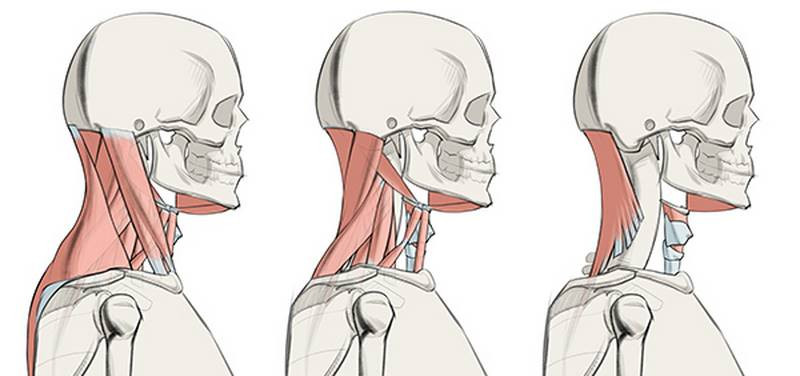
There are a lot of small muscles in the neck that pop out when the neck tenses and moves around. We’ll go over 10 muscles in the premium lesson. In this lesson, I’ll focus on the 2 muscles that affect the surface the most along with some bones and cartilage.
Bones and Cartilage
As always, anatomy starts with the bones. Note that the spine inserts on the back of the skull, completely behind the jaw. The throat sits in front of the spine, making up for half the width of the neck. The mandible, or jawbone, will be an important attachment point today. So will the “hook” of the mastoid process behind it. You can easily feel the mastoid process on your own head. It’s the hard, bony area on the base of the skull, just behind the bottom of your ear. Some neck muscles attach to the clavicles. Remember that there’s a small gap between the clavicles where the manubrium sits, about one eyeball wide, before they flow out into that Cupid’s bow shape.
Below we see the Adam’s apple aka the thyroid cartilage that surrounds and protects the voicebox. It sits just below the hyoid bone, in front of the spine and esophagus and all that but behind the muscular wall of the neck. The Adam’s apple is larger and has a 90 degree angle on men, but women actually have one too. “Eve’s apple” is just at a more open 120 degree angle so it’s less noticeable.
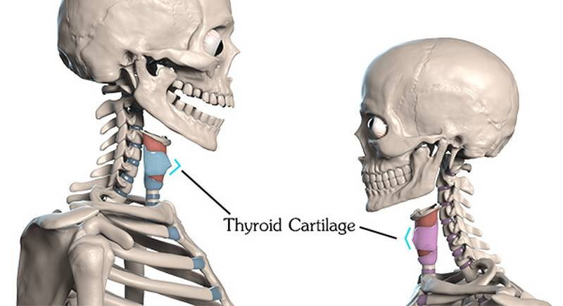
There’s another cartilage called the cricoid cartilage right under the thyroid cartilage
There’s a thyroid gland that cover the cricoid cartilage and soften it depending on how big the gland is.
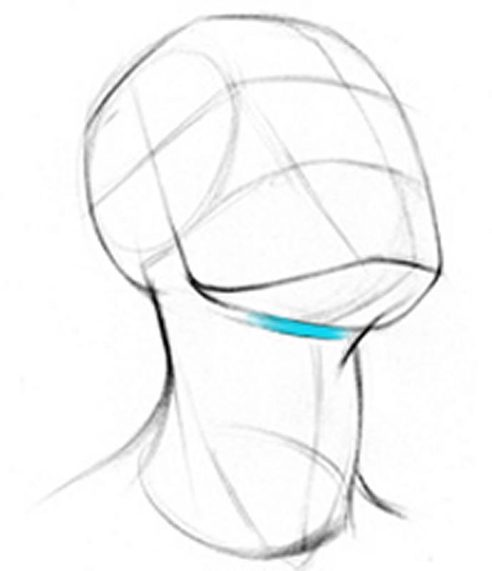
The hyoid is a small, horseshoe shaped bone above the Adam’s apple. It’s the corner between the bottom plane of the jaw and front of the neck. It’s an unusual little bone because it has no joints or direct attachments to other bones.
Time for muscles! There’s no competition: these three muscles are the largest and most important neck muscles for artists to know. The levator scapulae, sternocleidomastoideus, and trapezius are essential for expressing the forms and movements of the neck. We already covered the trapezius, so let’s get on with the other two!
Levator Scapulae
The levator scapulae is a diagonal muscle visible on the sides of the neck. It originates from the top 4 cervical vertebrae, and inserts on the top-most point of the scapula, at that medial-superior corner. The muscle twists on itself, so the fibers coming off of the highest point of the scapula attach the lowest on the neck.
The levator scapulae levitates the scapula, or rather, lifts up the medial edge. Sometimes it activates just to help stabilize the scapula, so you’ll see the levator scapulae popping out in a lot of different poses.
If you’re having trouble identifying neck muscles, the levator scapulae is the one that points to the ear. It’s buried under the sternomastoid anteriorly and by the trapezius posteriorly. But its middle third on the side of the neck is superficial.
Sternocleidomastoideus
It sounds intimidating, but the sternocleidomastoideus is probably a muscle you’re at least a little familiar with. It’s the one that makes the neck’s “V” shape as it goes from behind the ear to the pit of the neck. Its three-part name describes its three attachments... It originates from the top of sternum, "sterno", as well as the medial third of the clavicles, "cleido", and inserts on the mastoid process of the skull, "mastoideus". Sternocleidomastoideus! You can also call it sternomastoid for short.
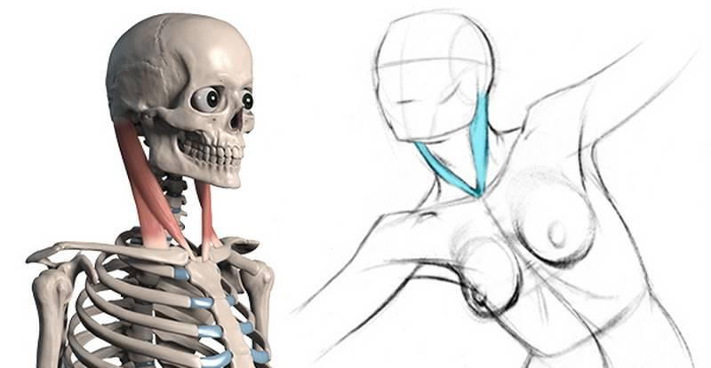
It has two distinct origins, and that means two distinct muscular heads, with a small gap above the clavicle. It’s superficial throughout its entire length, so the sternomastoid is a must-know muscle. When it activates it rotates the head to the opposite side. If both sides activate together, they flex the head forward. That’s the sternocleidomastoideus.
That's it for now. If you want to learn about 8 more neck muscles, check out the premium anatomy course.
